
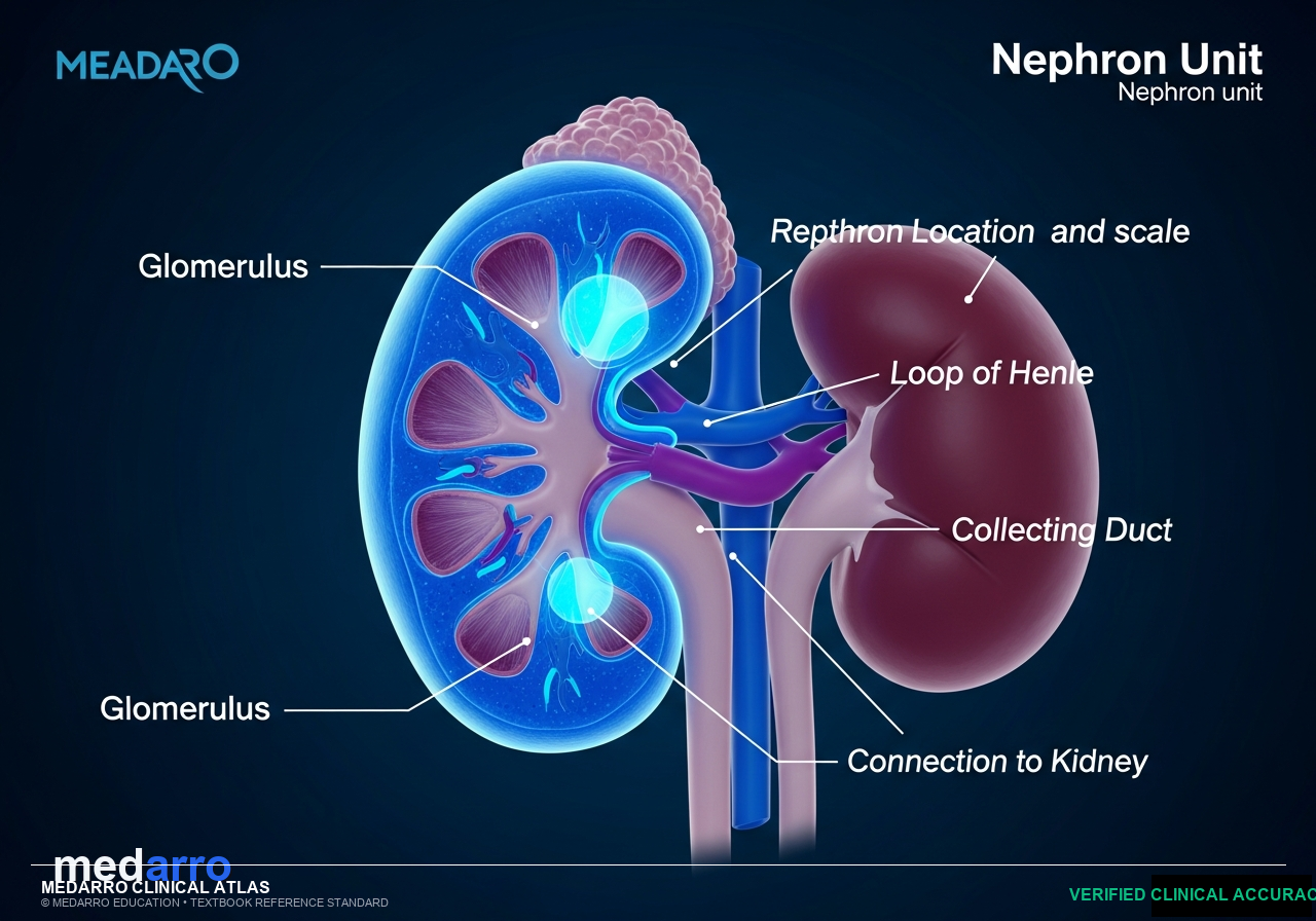
Nephron Histology: Glomerular Barrier & Countercurrent Transport
High-Yield Clinical Pearls & Exam Keys
The Proximal Convoluted Tubule (PCT) performs obligatory reabsorption of 65% of filtered water, sodium, chloride, and 100% of glucose and amino acids.
The Thick Ascending Limb (TAL) is completely impermeable to water, allowing active solute pumping via NKCC2 to generate the hypertonic medullary interstitial gradient.
Loss of negatively charged heparan sulfate proteoglycans in the glomerular basement membrane leads to massive selective albuminuria (Minimal Change Disease).
Ask Dr. Medarro about Human Anatomy
Instant NMC-grounded answers with standard textbook citations
Trilaminar Glomerular Filtration Barrier
Fenestrated Capillary Endothelium (70-90 nm pores)
Glomerular Basement Membrane with Type IV Collagen & Heparan Sulfate (Negative Charge)
Podocyte Foot Processes bridged by Nephrin & Podocin Slit Diaphragms (25-30 nm)
Tubular Segment Histology & Transport Characteristics
| Tubular Segment | Epithelial Lining | Primary Transporter & Target |
|---|---|---|
| Proximal Convoluted Tubule (PCT) | Simple tall cuboidal with dense microvilli brush border | SGLT2 (Glucose) & NHE3 (Na+/H+); 65% Na+/H2O reabsorbed |
| Thick Ascending Limb (TAL) | Simple cuboidal without brush border; water impermeable | NKCC2 symporter; generates medullary hypertonicity (Furosemide target) |
| Distal Convoluted Tubule (DCT) | Simple cuboidal without microvilli | NCCT symporter (Thiazide target); PTH-regulated Ca2+ reabsorption |
Interactive Clinical Vignette Practice
Live Rule-Out EngineA 4-year-old boy presents with sudden generalized periorbital and pretibial edema. Urinalysis shows 4+ proteinuria with normal renal function and no hematuria. Light microscopy of renal biopsy is normal. Electron microscopy reveals diffuse effacement of visceral epithelial foot processes. What component of the filtration barrier was primarily lost?
Unlock Complete Viva Traps & Additional Case Vignettes
Access model examiner answers, comprehensive rule-out breakdowns, and 4K spotter explanations.
Want 50+ Clinical Vignettes & 4K 3D Spotters on Human Anatomy?
Practice Spaced Repetition Decathlon, generate instant 7-day adaptive study plans, and upload your college PDFs into the personal vault.