Bailey & Love's Short Practice of Surgery 28th Ed · Ch 72 The Vermiform Appendix
Verified Clinical DiagramBailey & Love's Short Practice of Surgery 28th Ed · Ch 72 The Vermiform Appendix
High-Yield Clinical Pearls & Exam Keys
1
Luminal obstruction by a fecolith (appendicolith) in adults or lymphoid hyperplasia in children is the primary initiating event in acute appendicitis.
2
McBurney's point lies at the junction of the lateral one-third and medial two-thirds of a line connecting the right Anterior Superior Iliac Spine (ASIS) to the umbilicus.
3
Rovsing's sign is pain elicited in the right iliac fossa when deep pressure and palpation are applied to the left iliac fossa.
Ask Dr. Medarro about General Surgery
Instant NMC-grounded answers with standard textbook citations
AI Senior Mentor
Or tap a high-yield exam question:
Murphy's Triad of Acute Appendicitis
Key #1
Pain starting in the periumbilical region (visceral T10) and shifting to the right iliac fossa (somatic parietal peritoneum)
Key #2
Nausea and vomiting occurring AFTER the onset of pain
Key #3
Fever and localized tenderness over McBurney's point
Modified Alvarado Scoring System (MANTRELS)
Clinical Feature
Component Description
Points Allocated
M - Migration of Pain
Shift of pain from periumbilical to Right Iliac Fossa
1 Point
T - Tenderness in RIF
Localized physical tenderness at McBurney's point
2 Points (Double Weight)
L - Leukocytosis
White Blood Cell count > 10,000 /mm³
2 Points (Double Weight)
Interactive Clinical Vignette Practice
Live Rule-Out Engine
Case Vignette #1NEET-PG Calibrated
A 19-year-old male medical student presents with a 14-hour history of abdominal pain shifting from the periumbilicus to the right iliac fossa. What is the precise anatomical definition of McBurney's point?
AOne-third of the distance from the Right Anterior Superior Iliac Spine to the Umbilicus
BMidpoint of a line connecting the Umbilicus and the Symphysis Pubis
CTwo-thirds of the distance from the Right Anterior Superior Iliac Spine to the Umbilicus
DIntersection of the transumbilical plane and the midclavicular line
**1. Surgical Anatomy & Pathogenesis**
The vermiform appendix arises from the posteromedial wall of the cecum, approximately 2.5 cm inferior to the ileocecal valve, where the three taeniae coli converge. The most common anatomical position is **Retrocecal** (65–70%), followed by **Pelvic** (30%).
* **Primary Pathogenetic Event**: Luminal obstruction. In young adults, obstruction is caused by **lymphoid follicular hyperplasia** (often post-viral); in older adults, by a **fecolith** (appendicolith), foreign body, or carcinoid tumor.
* **Progression**: Continued mucus secretion behind the obstruction increases intraluminal pressure, compromising venous drainage, triggering bacterial invasion (*E. coli*, *Bacteroides fragilis*), ischemic mucosal necrosis, gangrene, and eventual perforation.
**2. Classical Symptom Progression (Murphy's Triad)**
Murphy's classical chronological sequence of symptoms occurs in over 80% of patients:
1. *Pain*: Begins as dull, poorly localized periumbilical visceral pain (referred along the T10 dermatome via the sympathetic lesser splanchnic nerves).
2. *Anorexia, Nausea, and Vomiting*: Anorexia is so consistent that hunger should prompt reconsideration of the diagnosis.
3. *Somatic Pain Shift*: Over 6 to 12 hours, transmural inflammation contacts the parietal peritoneum, causing the pain to shift and localize sharply to the **Right Iliac Fossa (RIF)**.
**3. Physical Signs on Examination**
* **McBurney's Point Tenderness**: Maximal tenderness located at the junction of the lateral 1/3 and medial 2/3 of a line connecting the anterior superior iliac spine (ASIS) to the umbilicus.
* **Rovsing's Sign**: Palpation of the left iliac fossa elicits pain in the right iliac fossa (due to retrograde displacement of colonic gas stretching the inflamed cecum/appendix).
* **Psoas Sign**: Passive hyperextension of the right hip with patient in left lateral position elicits pain, indicating an inflamed retrocecal appendix irritating the iliopsoas muscle.
* **Obturator Sign**: Pain elicited on passive internal rotation of the flexed right hip, indicating a pelvic appendix irritating the obturator internus muscle.
* **Blumberg's Sign**: Rebound tenderness indicating localized parietal peritonitis.
**4. 🔬 Diagnostic Scoring & Imaging**
* **Alvarado (MANTRELS) Score (Total = 10 Points)**:
- *Symptoms*: **M**igration of pain (1), **A**norexia (1), **N**ausea/Vomiting (1).
- *Signs*: **T**enderness in RIF (2), **R**ebound tenderness (1), **E**levated temperature $\ge$ 37.3°C (1).
- *Lab*: **L**eukocytosis >10,000/mcL (2), **S**hift to left of neutrophils (1).
- *Score $\ge$ 7*: High probability; strongly indicates need for emergency surgery.
* **Ultrasonography**: Graded-compression US shows a non-compressible, blind-ending, aperistaltic tubular structure with an outer diameter **>6 mm**, "target sign" on transverse view.
* **Contrast-Enhanced CT (CECT) of Abdomen**: Most sensitive (>95%) and specific imaging modality; demonstrates enlarged appendix (>6 mm), appendiceal wall thickening, periappendiceal fat stranding, and appendicoliths.
**5. Definitive Surgical Management**
* **Laparoscopic Appendectomy**: Gold standard. Advantages: reduced postoperative pain, lower wound infection rates, shorter hospital stay, and enables complete diagnostic peritoneal exploration.
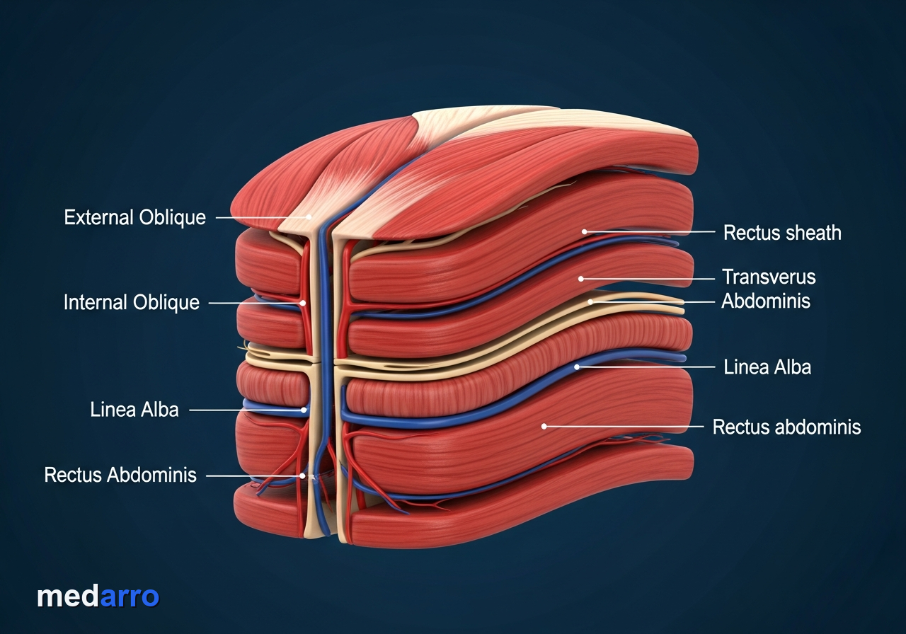
* **Open Appendectomy**: Performed via a transverse skin crease incision (Lanz) or gridiron muscle-splitting incision (McBurney's).
* **Antibiotic Prophylaxis**: Preoperative broad-spectrum IV Ceftriaxone (or Cefotaxime) plus Metronidazole administered within 60 minutes prior to incision.
**6. 📖 Standard Textbook Reference**
* *Bailey & Love's Short Practice of Surgery*, 28th Ed., Ch. 73: The Vermiform Appendix, pp. 1312–1326.
* *Sabiston Textbook of Surgery*, 21st Ed., Ch. 51: The Appendix, pp. 1295–1310.
Viva Examiner Traps & Model Answers
Oral Exam Prep
Why does the pain of acute appendicitis shift from the periumbilicus to the right iliac fossa?
Initial luminal distension stimulates visceral afferent C-fibers travelling to T10 spinal cord level (periumbilical). When inflammation breaches the serosa and irritates somatic parietal peritoneum, pain becomes sharply localized to the right iliac fossa via somatic intercostal nerves (T11-L1).
What is the artery of the appendix, and what is its surgical significance?
The Appendicular Artery, a branch of the inferior division of the ileocolic artery. It is an end-artery running in the free border of the mesoappendix; thrombosis or compression leads to rapid ischemic necrosis, gangrene, and perforation.
What ultrasonographic criteria confirm acute appendicitis?
A non-compressible, blind-ending tubular structure in the right lower quadrant with an outer diameter > 6 mm, 'target' sign in transverse section, appendicolith with acoustic shadowing, and periappendiceal fat stranding or fluid.
Additional Exam Practice Vignettes (1)
Case Vignette #2
A 24-year-old woman presents with right lower quadrant pain. During examination, deep pressure applied over the left iliac fossa reproduces sharp pain in the right lower quadrant. What is the name of this sign?
Rovsing's sign is positive when pressure on the left lower quadrant elicits pain in the right lower quadrant, caused by retrograde displacement of colonic gas irritating the inflamed right iliac fossa parietal peritoneum.
Free Student Account Required
Unlock Complete Viva Traps & Additional Case Vignettes
Access model examiner answers, comprehensive rule-out breakdowns, and 4K spotter explanations.